Powered by Blogger.

Wednesday, September 23, 2009

Appendicitis

What is Appendicitis?

Appendicitis is inflammation of the appendix, a small pouch attached to the cecum, the beginning of the colon, on the lower right side of the abdomen. The appendix is not necessary for life, but it can become diseased. If untreated, an inflamed appendix can burst, causing infection and even death. Appendicitis can affect people at any age. It is most common in people ages 10 to 30.

The cause of appendicitis is usually unknown. It may occur after a viral infection in the digestive tract or when the opening connecting the large intestine and appendix is blocked. The inflammation can cause infection, a blood clot, or rupture of the appendix. Because of the risk of rupture, appendicitis is considered an emergency. Anyone with symptoms needs to see a doctor immediately.

Symptoms of Appendicitis

The most common symptoms of appendicitis are:
pain in the right side of the abdomen
nausea
vomiting
constipation
diarrhea inability to pass gas
low fever that begins after other symptoms
abdominal swelling
anorexia

The pain usually begins near the navel and moves down and to the right. The pain becomes worse when moving, taking deep breaths, coughing, sneezing, and being touched in the area.

Not everyone with appendicitis has all the symptoms. People with symptoms of appendicitis should not take laxatives or enemas to relieve constipation because these medicines could cause the appendix to burst. Pain medicine can mask symptoms that the doctor needs to know about, so it should not be used before consulting a doctor when appendicitis is suspected.

The doctor bases an appendicitis diagnosis on symptoms, a physical exam, blood tests to check for signs of infection such as a high white blood cell count, and urine tests to rule out a urinary tract infection. Usually doctors use CT scan or ultrasound to see whether the appendix looks inflamed.

If the diagnosis of appendicitis is not certain, people with equivocal signs of appendicitis may be watched and sometimes treated with antibiotics. People with definite appendicitis have surgery to remove the appendix, which is called an appendectomy. Doctors may use laparoscopic surgery for appendectomy. This technique involves making several tiny cuts in the abdomen and inserting a miniature camera and surgical instruments. The surgeon then removes the appendix with the instruments, so there is usually no need to make a large incision in the abdomen. People can live a normal life without their appendix--changes in diet, exercise, or other lifestyle factors are not necessary.

Friday, September 18, 2009

Brest Health and Brest Cancer

The Breasts
Benign Breast Lumps
Breast Cancer Risk
Factors for Breast Cancer
Early Detection
Symptoms Diagnosis
Orthodox Treatment
Side Effects of Orthodox Treatment
The Breasts

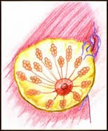
Each breast has 15 to 20 overlapping sections called lobes. Within each lobe are many smaller lobules, which end in dozens of tiny bulbs that can produce milk. The lobes, lobules, and bulbs are all linked by thin tubes called ducts. These ducts lead to the nipple in the center of a dark area of skin called the areola. Fat fills the spaces around the lobules and ducts. There are no muscles in the breast, but muscles lie under each breast and cover the ribs.Each breast also contains blood vessels and vessels that carry colorless fluid called lymph. The lymph vessels lead to small bean-shaped organs called lymph nodes. Clusters of lymph nodes are found near the breast in the axilla (under the arm), above the collarbone, and in the chest. Lymph nodes are also found in many other parts of the body.¡¡¡¡¡¡¡¡¡¡¡¡¡¡¡¡¡¡ TOP

Benign Breast Lumps
Any noticeable change, thickening or localized swelling in your breast that was not there before may be a lump. Eighty-five percent of all breast lumps found are benign. Some common benign breast problems that appear as lumps are:
Cystic Disease (fibrocystic breast disease) is the most common cause of breast lumps in women. These lumps or cysts are fluid-filled sacs that enlarge and become tender and painful before the menstrual cycle. This condition is responsible for at least half of all breast biopsies performed. It tends to involve both breasts. These lumps are movable, and if large may feel round and firm. Cystic disease usually disappears after menopause. Lipomas develop as single, painless lumps. They can vary in size. Lipomas consist of fatty tissue and are commonly found elsewhere in the body. Fibroadenomas, single solid tumors, appear most often in young women. These breast lumps are firm, rubbery, movable, often oval-shaped, and usually painless. Papillomas are small, wart-like growths in the lining of a mammary duct near the nipple. These can produce a bloody discharge of the nipple.
There are other, not as common benign breast lumps caused by a variety of breast problems such as sclerosing adenosis, etc. TOP

Breast Cancer

The most common type of breast cancer begins in the lining of the ducts and is called ductal carcinoma. Another type, called lobular carcinoma, arises in the lobules. When breast cancer spreads outside the breast, cancer cells are often found in the lymph nodes under the arm (axillary lymph nodes). If the cancer has reached these nodes, it may mean that cancer cells have spread to other parts of the body--other lymph nodes and other organs, such as the bones, liver, or lungs--via the lymphatic system or the bloodstream. Cancer that spreads is the same disease and has the same name as the original (primary) cancer. When breast cancer spreads, it is called metastatic breast cancer, even though the secondary tumor is in another organ. Doctors sometimes call this "distant" disease. TOP

Risk Factors for Breast Cancer

The risk of breast cancer increases gradually as a woman gets older. This disease is uncommon in women under the age of 35. All women age 40 and older are at risk for breast cancer. However, most breast cancers occur in women over the age of 50, and the risk is especially high for women over age 60. Research has shown that the following conditions place a woman at increased risk for breast cancer:

Personal history of breast cancer. Women who have had breast cancer face an increased risk of getting breast cancer again. Genetic alterations. Changes in certain genes (BRCA1, BRCA2, and others) make women more susceptible to breast cancer. Family history. A woman's risk for developing breast cancer increases if her mother, sister, daughter, or two or more other close relatives, such as cousins, have a history of breast cancer, especially at a young age. Certain breast changes. Having a diagnosis of atypical hyperplasia or lobular carcinoma in situ (LCIS) or having had two or more breast biopsies for other benign conditions may increase a woman's risk for developing cancer. Other factors associated with an increased risk for breast cancer include: 1. Breast density. Women age 45 and older whose mammograms show at least 75 percent dense tissue are at increased risk. 2. Radiation therapy. Women whose breasts were exposed to radiation during their childhood, especially those who were treated with radiation for Hodgkin's disease, are at an increased risk for developing breast cancer throughout their lives. 3. Late childbearing. Women who had their first child after the age of 30 have a greater chance of developing breast cancer than women who had their children at a younger age.
In most cases, doctors cannot explain why a woman develops breast cancer. Studies show that most women who develop breast cancer have none of the risk factors listed above, other than the risk that comes with growing older. Also, most women with known risk factors do not get breast cancer. Scientists are conducting research into the causes of breast cancer to learn more about risk factors and ways of preventing this disease.

Early Detection

When breast cancer is found and treated early, the chances for survival are better. Women can take an active part in the early detection of breast cancer by having regular screening mammograms and clinical breast exams (breast exams performed by health professionals). Some women also perform breast self-exams.
Symptoms

Early breast cancer usually does not cause pain. In fact, when breast cancer first develops, there may be no symptoms at all. But as the cancer grows, it can cause changes that women should watch for: A lump or thickening in or near the breast or in the underarm area; A change in the size or shape of the breast; Nipple discharge or tenderness, or the nipple pulled back (inversion) into the breast; Ridges or pitting of the breast (the skin looks like the skin of an orange; or A change in the way the skin of the breast, areola, or nipple looks or feels (for example, warm, swollen, red, or scaly). A woman should see her doctor about any symptoms like these. Most often, they are not cancer, but it's important to check with the doctor so that any problems can be diagnosed and treated as early as possible.

Diagnosis

In addition to checking general signs of health, a woman's doctor may do one or more of the breast exams described on the following page.
Palpation. The doctor can tell a lot about a lump (its size, its texture, and whether it moves easily) by palpation, carefully feeling the lump and the tissue around it. Benign lumps often feel different from cancerous ones. Mammography. X-rays of the breast can give the doctor important information about a breast lump. If an area on the mammogram looks suspicious or is not clear, additional mammograms may be needed. Ultrasonography. Using high-frequency sound waves, ultrasonography can often show whether a lump is solid or filled with fluid. This exam may be used along with mammography. Fine needle aspiration. A thin needle is used to remove fluid from a breast lump. This procedure may show whether a lump is a fluid-filled cyst (not cancer) or a solid mass (which may or may not be cancer). Clear fluid removed from a cyst may not need to be checked by a lab. Needle biopsy. Using special techniques, tissue can be removed with a needle from an area that is suspicious on a mammogram but cannot be felt. Tissue removed in a needle biopsy goes to a lab to be checked by a pathologist for cancer cells.Surgical biopsy. The surgeon cuts out part or all of a lump or suspicious area. A pathologist examines the tissue under a microscope to check for cancer cells. When cancer is found, the pathologist can tell what kind of cancer it is (whether it began in a duct or a lobule) and whether it is invasive (has invaded nearby tissues in the breast). Special lab tests of the tissue help the doctor learn more about the cancer. For example, hormone receptor tests (estrogen and progesterone receptor tests) can help predict whether the cancer is sensitive to hormones. TOP
Orthodox Treatment
The treatment options for each woman depend on the size and location of the tumor in her breast, the results of lab tests (including hormone receptor tests), and the stage (or extent) of the disease.

Methods of Treatment

Methods of treatment for breast cancer are local or systemic. Local treatments are used to remove, destroy, or control the cancer cells in a specific area. Surgery and radiation therapy are local treatments. Systemic treatments are used to destroy or control cancer cells throughout the body. Chemotherapy and hormonal therapy are systemic treatments. A patient may have just one form of treatment or a combination. Different forms of treatment may be given at the same time or one after another.
Surgery is the most common treatment for breast cancer. Several types of surgery may be used. An operation to remove the breast (or as much of the breast as possible) is a mastectomy. Breast reconstruction is often an option at the same time as the mastectomy, or later on. An operation to remove the cancer but not the breast is called breast-sparing surgery or breast-conserving surgery. Lumpectomy and segmental mastectomy (also called partial mastectomy) are types of breast-sparing surgery. They usually are followed by radiation therapy to destroy any cancer cells that may remain in the area.
In lumpectomy, the surgeon removes the breast cancer and some normal tissue around it. Often, some of the lymph nodes under the arm are removed.
In segmental mastectomy, the surgeon removes the cancer and a larger area of normal breast tissue around it. Occasionally, some of the lining over the chest muscles below the tumor is removed as well. Some of the lymph nodes under the arm may also be removed.
In total (simple) mastectomy, the surgeon removes the whole breast. Some of the lymph nodes under the arm may also be removed.
In modified radical mastectomy, the surgeon removes the whole breast, most of the lymph nodes under the arm, and often the lining over the chest muscles. The smaller of the two chest muscles is also taken out to help in removing the lymph nodes.
In radical mastectomy (also called Halsted radical mastectomy), the surgeon removes the breast, the chest muscles, all of the lymph nodes under the arm, and some additional fat and skin. Breast reconstruction (surgery to rebuild a breast's shape) is often an option after mastectomy.
Radiation therapy (also called radiotherapy) is the use of high-energy rays to kill cancer cells and stop them from growing. The rays may come from radioactive material outside the body and be directed at the breast by a machine (external radiation). The radiation can also come from radioactive material placed directly in the breast in thin plastic tubes (implant radiation). Some women receive both kinds of radiation therapy. Radiation therapy, alone or with chemotherapy or hormone therapy, is sometimes used before surgery to destroy cancer cells and shrink tumors. This approach is most often used in cases in which the breast tumor is large or not easily removed by surgery.
Chemotherapy is the use of drugs to kill cancer cells. Chemotherapy for breast cancer is usually a combination of drugs. The drugs may be given by mouth or by injection. Either way, chemotherapy is a systemic therapy because the drugs enter the bloodstream and travel throughout the body. Chemotherapy is given in cycles: a treatment period followed by a recovery period, then another treatment, and so on. Most patients have chemotherapy in an outpatient part of the hospital, at the doctor's office, or at home. Depending on which drugs are given and the woman's general health, however, she may need to stay in the hospital during her treatment.
Hormonal therapy is used to keep cancer cells from getting the hormones they need to grow. This treatment may include the use of drugs that change the way hormones work or surgery to remove the ovaries, which make female hormones. Like chemotherapy, hormonal therapy is a systemic treatment; it can affect cancer cells throughout the body.
Treatment decisions are complex. They are often affected by the judgment of the doctor, by the desires of the patient and, the most important, the stage of the disease. The stage is based on the size of the tumor and whether the cancer has spread. TOP
Side Effects of Orthodox Treatment
It is hard to limit the effects of cancer treatment so that only cancer cells are removed or destroyed. Because healthy cells and tissues may also be damaged, treatment often causes unwanted side effects. The side effects of cancer treatment are different for each person, and they may even be different from one treatment to the next.
Surgery Surgery causes short-term pain and tenderness in the area of the operation, so women may need to talk with their doctor about which method of pain control would be appropriate. Any kind of surgery also carries a risk of infection, poor wound healing, bleeding, or a reaction to the anesthesia used in surgery. Removal of a breast can cause a woman's weight to shift and be out of balance--especially if she has large breasts. This imbalance can cause discomfort in a woman's neck and back. Also, the skin in the breast area may be tight, and the muscles of the arm and shoulder may feel stiff. After a mastectomy, some women have some permanent loss of strength in these muscles. Because nerves may be injured or cut during surgery, a woman may have numbness and tingling in the chest, underarm, shoulder, and arm. These feelings usually go away within a few weeks or months, but some women may have permanent numbness. Removing the lymph nodes under the arm slows the flow of lymph. In some women, this fluid builds up in the arm and hand and causes swelling (lymphedema). Women need to protect the arm and hand on the treated side from injury, even long after surgery. The radiation may cause side effects that involve the heart, lungs, and ribs. One of the common side effects is fatigue, especially in the later weeks of treatment and for sometime afterward. It is also common for the skin in the treated area to become red, dry, tender, and itchy. Toward the end of treatment, the skin may become moist and "weepy." Exposing this area to air as much as possible will help the skin heal. Because bras and some types of clothing may rub the skin and cause irritation, patients may want to wear loose-fitting cotton clothes. These effects of radiation therapy on the skin are temporary, and the area gradually heals once treatment is over. However, there may be a permanent change in the color of the skin. For most women, the breast will look and feel about the same after radiation therapy. Occasionally, the treated breast may be firmer. Also, it may be larger (due to fluid buildup) or smaller (because of tissue changes) than it was before. For some women, the breast skin is more sensitive after radiation treatment; for others, it is less sensitive.
Chemotherapy The side effects of chemotherapy depend mainly on the drugs the patient receives. As with other types of treatment, side effects vary from person to person. In general, anticancer drugs affect rapidly dividing cells. These include blood cells, which fight infection, cause the blood to clot, and carry oxygen to all parts of the body. When blood cells are affected by anticancer drugs, patients are more likely to get infections, bruise or bleed easily, and may have less energy during treatment and for some time afterward. Cells in hair follicles and cells that line the digestive tract also divide rapidly. As a result of chemotherapy, patients may lose their hair and may have other side effects, such as loss of appetite, nausea, vomiting, diarrhea, or mouth sores. There are cases in which the heart is weakened, and second cancers such as leukemia (cancer of the blood cells) have occurred. Also, some anticancer drugs can damage the ovaries. If the ovaries fail to produce hormones, the woman may have symptoms of menopause, such as hot flashes and vaginal dryness. Her periods may become irregular or may stop, and she may not be able to become pregnant. However, some women may still be able to get pregnant during treatment. Because the effects of chemotherapy on an unborn child are not known, it is important for a woman to talk to her doctor about birth control before treatment begins. After treatment, some women regain their ability to become pregnant, but in women over the age of 35 or 40, infertility is likely to be permanent.
Hormonal Therapy Hormonal therapy can cause a number of side effects. They depend largely on the specific drug or type of treatment, and they vary from patient to patient. Tamoxifen is the most common hormonal treatment. This drug blocks the body's use of estrogen but does not stop estrogen production. Tamoxifen may cause hot flashes, vaginal discharge or irritation, and irregular periods. Any unusual bleeding should be reported to the doctor. Younger women taking tamoxifen may become pregnant more easily and should discuss birth control methods with their doctor. Serious side effects of tamoxifen are rare, but this drug can cause blood clots in the veins, especially in the legs. In a very small number of women, tamoxifen has caused cancer of the lining of the uterus. The doctor may do a pelvic exam, as well as biopsies or other tests of the lining of the uterus, to monitor for this condition. (This does not apply to women who have had a hysterectomy, surgery to remove the uterus.) Young women whose ovaries are removed to deprive the cancer cells of estrogen experience menopause immediately. The side effects they have are likely to be more severe than the effects of natural menopause.

Wednesday, September 16, 2009

What is MRI and how does it work?

What is MRI and how does it work?
Magnetic resonance imaging, or MRI, is a way of obtaining very detailed images of organs and tissues throughout the body without the need for x-rays. Instead, MRI uses a powerful magnetic field, radio waves, a rapidly changing magnetic field, and a computer to create images that show whether or not there is an injury or some disease process present. For this procedure, the patient is placed within the MR scanner—typically a large, tunnel or doughnut-shaped magnet that is open at both ends. The magnetic field aligns atomic particles called protons that are present in most of the body's tissues. Radio waves then cause these particles to produce signals that are picked up by a receiver within the MR scanner. The signals are specially characterized using a changing magnetic field, and computer-processor to create very sharp images of tissues as "slices" that can be viewed in any orientation.
An MRI exam causes no pain, and the magnetic fields produce no known tissue damage of any kind. The MR scanner may make loud tapping or knocking noises at times during the exam; using earplugs prevents problems that may occur with this noise. You will be able to communicate with the MRI technologist or radiologist at any time using an intercom system or by other means. What is MRI used for?
MRI has become the preferred procedure for diagnosing a large number of potential problems or abnormal conditions in many different parts of the body. In general, MRI creates pictures that can show differences between healthy and unhealthy tissues. Physicians use MRI to examine the brain, spine, joints (e.g., knee, shoulder, hip, wrist, and ankle), abdomen, pelvic region, breast, blood vessels, heart and other body parts.How safe is MRI?
The powerful magnetic field of the MR system will attract iron-containing (also known as ferromagnetic) objects and may cause them to move suddenly and with great force. This can pose a possible risk to the patient or anyone in an object's "flight path." Great care is taken to be certain that objects such as "ferromagnetic" screwdrivers and oxygen tanks are not brought into the MR system area. It is vital that you remove all metallic belongings in advance of an MRI exam, including watches, jewelry, and items of clothing that have metallic threads or fasteners.
The powerful magnetic field of the MR system will pull on any iron-containing object in the body, such as certain medication pumps or aneurysm clips. Every MRI facility has a screening procedure and protocol that, when carefully followed, ensures that the MRI technologist and radiologist knows about the presence of metallic implants and materials so that special precautions can be taken. In some unusual cases the exam may have to be canceled. For example, the MRI exam will not be performed if a "ferromagnetic" aneurysm clip is present, because there is a risk of the clip moving or being dislodged. In some cases, certain medical implants can heat substantially during the MRI examination. Therefore, it is very important to inform the MRI technologist about any implant or other internal object that you may have.
The magnetic field of the MR systems may damage an external hearing aid or cause a heart pacemaker or electrical stimulator, or neurostimulator, to malfunction or cause patient injury. If you have a bullet or other metallic fragment in your body there is a potential risk that it could change position, possibly causing injury.
In addition, a metallic implant or other object may cause signal loss or distort the MR images. This may be unavoidable, but if the radiologist knows about it, allowances can be made when interpreting the MR images.
For some MRI studies a contrast material called "gadolinium" may be injected into a vein to help improve the information seen on the MR images. Unlike contrast agents used in x-ray studies, a gadolinium-based contrast agent does not contain iodine and, therefore, rarely causes an allergic reaction or other problem. However, if you have a history of kidney disease, kidney failure, kidney transplant, or liver disease, you must inform the MRI technologist and/or radiologists before receiving a gadolinium-based contrast agent. If you are unsure about the presence of these conditions, please discuss these matters with the technologist or radiologist.How should I prepare for my MRI exam?
You will typically receive a gown to wear during your MRI examination. Before entering the MR system room, you and any accompanying friend or relative will be asked questions regarding the presence of implants and will be instructed to remove all metal objects from pockets and hair. Additionally, the accompanying individual will need to fill out a screening form to ensure that he or she may safely enter the MR system room.
Before the exam you will be asked to fill out a screening form asking about anything that might create a health risk or interfere with imaging. Items that may create a health hazard or other problem during an MRI exam include:Cardiac pacemaker or implantable defibrillatorCatheter that has metal components that may pose a risk of a burn injuryA ferromagnetic metal clip placed to prevent bleeding from an intracranial aneurysmAn implanted or external medication pump (such as that used to deliver insulin or a pain-relieving drug)A cochlear (inner ear) implant
Items that need to be removed by patients and individuals before entering the MR system room include:Purse, wallet, money clip, credit cards, cards with magnetic stripsElectronic devices such as beepers or cell phonesHearing aidsMetal jewelry, watchesPens, paper clips, keys, coinsHair barrettes, hairpinsAny article of clothing that has a metal zipper, buttons, snaps, hooks, underwires, or metal threadsShoes, belt buckles, safety pins
Objects that may interfere with image quality if close to the area being scanned include:Metallic spinal rodPlates, pins, screws, or metal mesh used to repair a bone or jointJoint replacement or prosthesisMetal jewelry including those used for body piercingSome tattoos or tattooed eyeliner (these alter MR images, and there is a chance of skin irritation or swelling; black and blue pigments are the most troublesome)Bullet, shrapnel, or other type of metal fragmentMetallic foreign body within or near the eye (such an object generally can be seen on an x-ray; metal workers are most likely to have this problem)Dental fillings (while usually unaffected by the magnetic field, they may distort images of the facial area or brain; the same is true for orthodontic braces and retainers)An example of the MRI examination.
The MRI examination is performed in a special room that houses the MR system or "scanner." You will be escorted into the room by a staff member of the MRI facility and asked to lie down on a comfortably padded table that gently glides you into the scanner.
In general, in preparation for the MRI examination, you may be required to wear earplugs or headphones to protect your hearing because, when certain scanners operate, they may produce loud noises. These loud noises are normal and should not worry you.
For some MRI studies, a contrast agent called "gadolinium" may be injected into a vein to help obtain a clearer picture of the area being examined. At some point during the examination, a nurse or technologist will slide the table out of the scanner in order to inject the contrast agent. This is typically done through a small needle connected to an intravenous line that is placed in an arm or hand vein. A saline solution will drip through the intravenous line to prevent clotting until the contrast material is injected at some point during the exam.
The most important thing for the patient to do is to relax and lie still. Most MRI exams take between 15 to 45 minutes to complete depending on the body part imaged and how many images are needed, although some may take as long as 60 minutes or longer. You will be told ahead of time how long your scan is expected to take.
You will be asked to remain perfectly still during the time the imaging takes place, but between sequences some minor movement may be allowed. The MRI technologist will advise you accordingly.
When the MRI procedure begins, you may breathe normally, however, for certain examinations it may be necessary for you to hold your breath for a short period of time.
During your MRI examination, the MR system operator will be able to speak to you, hear you, and observe you at all times. Consult the scanner operator if you have any questions or feel anything unusual.
When the MRI procedure is over, you may be asked to wait until the images are examined to determine if more images are needed. After the scan, you have no restrictions and can go about your normal activities.
Once the entire MRI examination is completed, the images will be looked at by a radiologist, a specially-trained physician who is able to interpret the scans for your doctor. The question of claustrophobia
Some patients who have MRI may feel confined, closed-in, or frightened. Perhaps one in twenty will require a sedative to remain calm. Today, many patients avoid this problem when examined in one of the newer MRI units that have a more "open" design. Some MRI centers permit a relative or friend to be present in the MR system room, which also has a calming effect. If patients are properly prepared and know what to expect, it is almost always possible to complete the examination. Pregnancy and MRI
If you are pregnant or suspect you are pregnant, you should inform the MRI technologist and/or radiologist during the screening procedure. In general, there is no known risk of using MRI in pregnant patients. However, MRI is reserved for use in pregnant patients only to address very important problems or suspected abnormalities. In any case, MRI is safer for the fetus than imaging with x-rays.
You should inform your radiologist if you are breast-feeding at the time of a scheduled MRI study and may need to receive an MRI contrast agent. One option under this circumstance is to pump breast milk before the study, to be used until injected contrast material has cleared from the body, which typically takes about 24 hours.

Brest Health :MRI-Guided Breast Biopsy

MRI-Guided Breast Biopsy
What is MR-Guided Breast Biopsy?

Lumps or abnormalities in the breast are often detected by physical examination, mammography, or other imaging studies. However, it is not always possible to tell from these imaging tests whether a growth is benign or cancerous.
A breast biopsy is performed to remove some cells—either surgically or through a less invasive procedure involving a hollow needle—from a suspicious area in the breast and examine them under a microscope to determine a diagnosis. Image-guided needle biopsy is not designed to remove the entire lesion, but most of a very small lesion may be removed in the process of biopsy.
Image-guided biopsy is performed when the abnormal area in the breast is too small to be felt, making it difficult to locate the lesion by hand (called palpation).
In MRI-guided breast biopsy, magnetic resonance imaging is used to help guide the radiologist's instruments to the site of the abnormal growth.
top of pageWhat are some common uses of the procedure?
An MRI-guided breast biopsy is most helpful when MR imaging shows a breast abnormality such as:a suspicious mass not identified by other imaging techniquesan area of distortionan area of abnormal tissue change
MRI guidance is used in four biopsy procedures:fine needle aspiration (FNA), which uses a very small needle to extract fluid or cells from the abnormal area.core needle (CN) which uses a large hollow needle to remove one sample of breast tissue per insertion.vacuum-assisted device (VAD) which uses a vacuum powered instrument to collect multiple tissue samples during one needle insertion.wire localization, in which a guide wire is placed into the suspicious area to help the surgeon locate the lesion for surgical biopsy.


How should I prepare?

You may be asked to wear a gown during the exam or you may be allowed to wear your own clothing if it is loose-fitting and has no metal fasteners.
Guidelines about eating and drinking before an MRI exam vary with the specific exam and also with the facility. For some types of exams, you will be asked to fast for 8-12 hours. Unless you are told otherwise, you may follow your regular daily routine and take medications as usual.
Some MRI examinations may require the patient to swallow contrast material or receive an injection of contrast into the bloodstream. The radiologist or technologist may ask if you have allergies of any kind, such as allergy to iodine or x-ray contrast material, drugs, food, the environment, or asthma. However, the contrast material used for an MRI exam, called gadolinium, does not contain iodine and is less likely to cause side effects or an allergic reaction.
The radiologist should also know if you have any serious health problems or if you have recently had surgery. Some conditions, such as severe kidney disease may prevent you from being given contrast material for an MRI.
Women should always inform their physician or technologist if there is any possibility that they are pregnant. MRI has been used for scanning patients since the 1980's with no reports of any ill effects on pregnant women or their babies. However, because the baby will be in a strong magnetic field, pregnant women should not have this exam unless the potential benefit from the MRI is assumed to outweigh the potential risks. See the Safety page for more information about pregnancy and MR imaging.
Prior to a needle biopsy, you should report to your doctor all medications that you are taking, including herbal supplements, and if you have any allergies, especially to anesthesia. Your physician will advise you to stop taking aspirin or a blood thinner three days before your procedure.
Also, inform your doctor about recent illnesses or other medical conditions.
You may want to have a relative or friend accompany you and drive you home afterward. This is recommended if you have been sedated.
There are other important guidelines for patients to follow prior to undergoing MR imaging. For a list of these and a review of all preparations that should be made prior to MR imaging, please see MRI of the Breast.

What does the equipment look like?


>The traditional MRI unit is a large cylinder-shaped tube surrounded by a circular magnet. You will lie on a moveable examination table that slides into the center of the magnet.
Some MRI units, called short-bore systems, are designed so that the magnet does not completely surround you; others are open on the sides ("low-strength" open MRI). These units are especially helpful for examining patients who are fearful of being in a closed space and for those who are very obese. Newer open MRI units provide very high quality images for many types of exams; however, open MRI units with older magnets may not provide this same quality. Certain types of exams cannot be performed using open MRI. For more information, consult your doctor.
The computer workstation that processes the imaging information is located in a separate room than the scanner.
The majority of MRI-guided breast biopsies are currently performed in closed MRI systems with a specially modified exam table. This moveable examination table allows your breasts to hang freely into cushioned openings, which contain wire coils that send and receive radio waves to help create the MR images.
One of four instruments will be used: A fine needle attached to a syringe, smaller than needles typically used to draw blood.A core needle, also called an automatic, spring-loaded needle, which consists of an inner needle connected to a trough, or shallow receptacle, covered by a sheath and attached to a spring-loaded mechanism. A vacuum-assisted device (VAD), a vacuum-powered instrument that uses pressure to pull tissue into the needle.A thin guide wire, which is used for a surgical biopsy.
Other sterile equipment involved in this procedure includes syringes, sponges, forceps, scalpels and a specimen cup or microscope slide.


top of pageHow does the procedure work?


Unlike conventional x-ray examinations and computed tomography (CT) scans, MRI does not depend on ionizing radiation. Instead, while in the magnet, radio waves redirect the axes of spinning protons, which are the nuclei of hydrogen atoms, in a strong magnetic field.
The magnetic field is produced by passing an electric current through wire coils in most MRI units. Other coils, located in the machine and in some cases, placed around the part of the body being imaged, send and receive radio waves, producing signals that are detected by the coils.
A computer then processes the signals and generates a series of images each of which shows a thin slice of the body. The images can then be studied from different angles by the interpreting physician.
Overall, the differentiation of abnormal (diseased) tissue from normal tissues is often better with MRI than with other imaging modalities such as x-ray, CT and ultrasound.
Using MRI guidance to calculate the position of the abnormal tissue and to verify the placement of the needle, the radiologist inserts the biopsy needle through the skin, advances it into the lesion and removes tissue samples. If a surgical biopsy is being performed, MRI may be used to guide a wire into the mass to help the surgeon locate the area for excision.
top of pageHow is the procedure performed?
Image-guided, minimally invasive procedures such as MR-guided breast biopsies are most often performed by a specially trained breast radiologist.
Breast biopsies are usually done on an outpatient basis.
You will lie face down on a moveable exam table and the affected breast or breasts will be positioned into openings in the table.
A nurse or technologist will insert an intravenous (IV) line into a vein in your hand or arm and the contrast material gadolinium will be given intravenously.
Your breast will be gently compressed between two compression plates (similar to those used in a diagnostic MRI exam), one of which is marked with a grid structure. Using computer software, the radiologist measures the position of the lesion with respect to the grid and calculates the position and depth of the needle placement.
A local anesthetic will be injected into the breast to numb it.
A very small nick is made in the skin at the site where the biopsy needle is to be inserted.
The radiologist then inserts the needle, advances it to the location of the abnormality and MR imaging is performed to verify its position. Depending on the type of MRI unit being used, you may remain in place or be moved out of the center or bore of the MRI scanner.
Tissue samples are then removed using one of three methods:In a fine needle aspiration, a fine gauge needle and a syringe withdraw fluid or clusters of cells.In a core needle biopsy, the automated mechanism is activated, moving the needle forward and filling the needle trough, or shallow receptacle, with 'cores' of breast tissue. The outer sheath instantly moves forward to cut the tissue and keep it in the trough. This process is repeated three to six times.With a vacuum-assisted device (VAD), vacuum pressure is used to pull tissue from the breast through the needle into the sampling chamber. Without withdrawing and reinserting the needle, it rotates positions and collects additional samples. Typically, eight to 10 samples of tissue are collected from around the lesion.


After this sampling, the needle will be removed


.
If a surgical biopsy is being performed, a wire is inserted into the suspicious area as a guide for the surgeon.
A small marker may be placed at the site so that it can be located in the future if necessary.
Once the biopsy is complete, pressure will be applied to stop any bleeding and the opening in the skin is covered with a dressing. No sutures are needed.
A mammogram may be performed to confirm that the marker is in the proper position.
This procedure is usually completed within 45 minutes.
top of pageWhat will I experience during and after the procedure?
You will be awake during your biopsy and should have little or no discomfort. Most women report little or no pain and no scarring on the breast.
Some women find that the major discomfort of the procedure is from lying on their stomach for the length of the procedure, which can be reduced by strategically placed cushions.
When you receive the local anesthetic to numb the skin, you will feel a slight pin prick from the needle. You may feel some pressure when the biopsy needle is inserted.
The area will become numb within a short time.
You must remain still while the biopsy is performed.
As tissue samples are taken, you may hear clicks from the sampling instrument.
If you experience swelling and bruising following your biopsy, you may be instructed to take an over-the-counter pain reliever and to use a cold pack. Temporary bruising is normal.
You should contact your physician if you experience excessive swelling, bleeding, drainage, redness or heat in the breast.
If a marker is left inside the breast to mark the location of the biopsied lesion, it will cause no pain, disfigurement or harm.
You should avoid strenuous activity for 24 hours after returning home, but then usually will be able to resume normal activities.
It is recommended that nursing mothers not breastfeed for 36 to 48 hours after an MRI in which a contrast material was given.


top of pageWho interprets the results and how do I get them?


A pathologist examines the removed specimen and makes a final diagnosis. Depending on the facility, the radiologist or your referring physician will share the results with you.
top of pageWhat are the benefits vs. risks?BenefitsThe procedure is less invasive than surgical biopsy, leaves little or no scarring and can be performed in less than an hour.MRI is a noninvasive imaging technique that does not involve exposure to ionizing radiation.MRI-guided breast biopsy using a core needle is considered both safe and accurate.The speed, accuracy and safety of MRI-guided vacuum-assisted breast biopsy are as good as MR-guided wire localization without the associated complications and cost of surgery.Compared with stereotactic biopsy, the MRI-guided method avoids the need for ionizing radiation exposure.MRI-guided breast biopsy, using either the core needle method or the vacuum-assisted device, takes less time than surgical biopsy, causes less tissue damage, and is less costly.Recovery time is brief and patients can soon resume their usual activities. RisksBecause the vacuum-assisted device removes slightly larger pieces of tissue than other types of needles, there is a risk of bleeding and forming a hematoma, or a collection of blood at the biopsy site. The risk, however, appears to be less than one percent of patients.An occasional patient has significant discomfort, which can be readily controlled by non-prescription pain medication.Any procedure where the skin is penetrated carries a risk of infection. The chance of infection requiring antibiotic treatment appears to be less than one in 1,000. Doing a biopsy of tissue located deep within the breast carries a slight risk that the needle will pass through the chest wall, allowing air around the lung that could collapse a lung. This is a rare occurrence.


What are the limitations of MRI-Guided Breast Biopsy?


MRI-guided tissue sampling is limited by the position of the abnormality in the breast. Breast lesions located in the back of the breast or small abnormalities can be difficult or impossible to accurately target using MR.
Breast biopsy procedures will occasionally miss a lesion or underestimate the extent of disease present. If the diagnosis remains uncertain after a technically successful procedure, surgical biopsy will usually be necessary.
The MR-guided breast biopsy method cannot be used unless the mass can be seen on an MRI exam. Calcifications within a cancerous nodule are not shown as clearly with MR as with x-rays. Small lesions may be difficult to target accurately by MR-guided breast biopsy.
The widespread use of this technique is limited by its high cost, availability, and length of the procedure. MR-guided biopsy should not be considered if the lesion can be seen on mammography or on ultrasound, where the biopsy can be performed more easily with less patient discomfort. In those cases, stereotactic biopsy or ultrasound-guided biopsy are the more appropriate methods of tissue sampling.

Sunday, September 6, 2009

Bra History

Bra History

Women have used garments designed to lift, separate and restrain their breast since as early as 2000 BC. From the 1500s until the 1800s the corset was the primary under-garment used by women for the purpose of shaping the waist and lifting the breasts.

In 1893, Marie Tucek patented the “Breast Supporter” – the first garment similar to the modern-day bra that used shoulder straps with a hook-and-eye closure to support the breasts in pockets of fabric. By 1907, the term “brassiere” began to show up in high-profile women's magazines and eventually, around 1912, it appeared in the Oxford English Dictionary.

In 1913, New York socialite Mary Phelps Jacob took matters into her own hands and fashioned her own corset replacement out of two silk handkerchiefs and some ribbon. Her friends were sold on this innovative idea and encouraged Mary to apply for a patent for her “Backless Brassiere” design. Within a short time, Mary lost interest in the garment business and sold her patent to Warner Brothers Corset Company for $1,500. Today, Warner Brothers is a leading name brand manufacturer of bras. Check out the extensive selection of bras on our bra styles page.

Saturday, September 5, 2009

Anatomy and Function of Brest (Lymphatic drainage,Shape and support,Development,Changes, Breastfeeding,Sexual role,Other suggested functions)


Anatomy 

Breast schematic diagram (adult female human cross section) - Legend: 1. Chest wall 2. Pectoralis muscles 3. Lobules 4. Nipple 5. Areola 6. Duct 7. Fatty tissue 8. Skin

Breasts are modified sudoriferous (sweat) glands which produce milk in women, and in some rare cases, in men.[2] Each breast has one nipple surrounded by the areola. The areola is colored from pink to dark brown and has several sebaceous glands. In women, the larger mammary glands within the breast produce the milk. They are distributed throughout the breast, with two-thirds of the tissue found within 30 mm of the base of the nipple.[3] These are drained to the nipple by between 4 and 18 lactiferous ducts, where each duct has its own opening. The network formed by these ducts is complex, like the tangled roots of a tree. It is not always arranged radially, and branches close to the nipple. The ducts near the nipple do not act as milk.reservoirs; Ramsay et al. have shown that conventionally described lactiferous sinuses do not, in fact, exist. Instead, most milk is actually in the back of the breast, and when suckling occurs, the smooth muscles of the gland push more milk forward.

The remainder of the breast is composed of connective tissue (collagen and elastin), adipose tissue (fat), and Cooper's ligaments. The ratio of glands to adipose tissues rises from 1:1 in nonlactating women to 2:1 in lactating women.[3]

The breasts sit over the pectoralis major muscle and usually extend from the level of the 2nd rib to the level of the 6th rib anteriorly. The superior lateral quadrant of the breast extends diagonally upwards towards the axillae and is known as the tail of Spence. A thin layer of mammary tissue extends from the clavicle above to the seventh or eighth ribs below and from the midline to the edge of the latissimus dorsi posteriorly. (For further explanation, see anatomical terms of location.)

The arterial blood supply to the breasts is derived from the internal thoracic artery (formerly called the internal mammary artery), lateral thoracic artery, thoracoacromial artery, and posterior intercostal arteries. The venous drainage of the breast is mainly to the axillary vein, but there is some drainage to the internal thoracic vein and the intercostal veins. Both sexes have a large concentration of blood vessels and nerves in their nipples. The nipples of both women and men can become erect in response to sexual stimuli,[4] and also to cold.

The breast is innervated by the anterior and lateral cutaneous branches of the fourth through sixth intercostal nerves. The nipple is supplied by the T4 dermatome.

Lymphatic drainage


About 75% of lymph from the breast travels to the ipsilateral axillary lymph nodes. The rest travels to parasternal nodes, to the other breast, or abdominal lymph nodes. The axillary nodes include the pectoral, subscapular, and humeral groups of lymph nodes. These drain to the central axillary lymph nodes, then to the apical axillary lymph nodes. The lymphatic drainage of the breasts is particularly relevant to oncology, since breast cancer is a common cancer and cancer cells can break away from a tumour and spread to other parts of the body through the lymph system by metastasis.

Shape and support

Breasts vary in size, shape and position on a woman's chest, and their external appearance is not predictive of their internal anatomy or lactation potential. The natural shape of a woman's breasts is primarily dependent on the support provided by the Cooper's ligaments and the underlying chest on which they rest (the base). The breast is attached at its base to the chest wall by the deep fascia over the pectoral muscles. On its upper surface it is given some support by the covering skin where it continues on to the upper chest wall. It is this support which determines the shape of the breasts. In a small number of women, the frontal ducts (ampullae) in the breasts are not flush with the surrounding breast tissue, which causes the sinus area to visibly bulge outward.


Relatively round breasts which protrude almost horizontally.

Some breasts are high and rounded, and protrude almost horizontally from the chest wall. Such high breasts are common for girls and women in early stages of development. The protruding or high breasts are anchored to the chest at the base, and the weight is distributed evenly over the area of the base of the approximately dome- or cone-shaped breasts.[citation needed]

In the “low” breast, a proportion of the breasts' weight is actually supported by the chest against which the lower breast surface comes to rest, as well as the deep anchorage at the base. The weight is thus distributed over a larger area, which has the effect of reducing the strain. In both males and females, the thoracic cavity slopes progressively outwards from the thoracic inlet (at the top of the breastbone) above to the lowest ribs which mark its lower boundary, allowing it to support the breasts.

The inframammary fold (or line, or crease) is an anatomic structure created by adherence between elements in the skin and underlying connective tissue[5] and represents the inferior extent of breast anatomy. Some teenagers may develop breasts whose skin comes into contact with the chest below the fold at an early age, and some women may never develop such breasts; both situations are perfectly normal. The relationship of the nipple position to the fold is described as ptosis, a term also applied to other body parts and which refers in general to drooping or sagging. Due to breast weight and relaxation of support structures, the nipple-areola complex and breast tissue may eventually hang below the fold, and in some cases the breasts may extend as far as, or even beyond, the navel. The length from the nipple to the sternal notch (central, upper border) in the youthful breast averages 21 cm and is a common anthropometric figure used to assess both breast symmetry and ptosis. Lengthening of both this measurement and the distance between the nipple and the fold are both characteristic of advancing grades of ptosis.

The end of the breast, which includes the nipple, may either be flat (a 180° angle) or angled (angles lower than 180°). Breast ends are rarely angled sharper than 60°. Angling of the end of the breast is caused in part by the ligaments that suspend it, such that the breast ends often have a more obtuse angle when a woman is lying on her back. Breasts exist in a range of ratios between length and base diameter, usually ranging from ½ to 1.

Development
 
Male with severe gynecomastia

Girls develop breasts during puberty, as a result of changing sex hormones, chiefly estrogen, which also has been demonstrated to cause the development of woman-like, enlarged breasts in men, a condition called gynecomastia.

In most cases, the breasts fold down over the chest wall during Tanner stage development, as shown in this diagram.[6] It is typical for a woman's breasts to be unequal in size particularly while the breasts are developing. Statistically it is slightly more common for the left breast to be the larger.[7] In rare cases, the breasts may be significantly different in size, or one breast may fail to develop entirely.

A large number of medical conditions are known to cause abnormal development of the breasts during puberty. Virginal breast hypertrophy is a condition which involves excessive growth of the breasts, and in some cases the continued growth beyond the usual pubescent age. Breast hypoplasia is a condition where one or both breasts fail to develop.

Changes

Breast with visible stretchmarks.

As breasts are mostly composed of adipose tissue, their size can change over time. This occurs for a number of reasons, most obviously when a girl grows during puberty and when a woman becomes pregnant. The breast size may also change if she gains (or loses) weight for any other reason. Any rapid increase in size of the breasts can result in the appearance of stretchmarks.


It is typical for a number of other changes to occur during pregnancy: in addition to becoming larger, the breasts generally become firmer, mainly due to hypertrophy of the mammary gland in response to the hormone prolactin. The size of the nipples may increase noticeably and their pigmentation may become darker. These changes may continue during breastfeeding. The breasts generally revert to approximately their previous size after pregnancy, although there may be some increased sagging and stretchmarks.

The size of a woman's breasts may fluctuate during the menstrual cycle, particularly with premenstrual water retention. An increase in breast size is a common side effect of use of the combined oral contraceptive pill.

Breasts sag if the ligaments become elongated, a natural process that can occur over time and is also influenced by the breast bouncing while exercising. Breasts can decrease in size at menopause if estrogen levels decline.

Function of Brest  

Breastfeeding


An infant breastfeeding
Main article: Breastfeeding


The primary function of mammary glands is to nurture young by producing breast milk. The production of milk is called lactation. (While the mammary glands that produce milk are present in the male, they normally remain undeveloped.) The orb-like shape of breasts may help limit heat loss, as a fairly high temperature is required for the production of milk. Alternatively, one theory states that the shape of the human breast evolved in order to prevent infants from suffocating while feeding.[8] Since human infants have a small jaw (not protruding, like other primates), the infant's nose might be blocked if the mother's chest was too flat.[8] According to this theory, as the human jaw receded, the breasts became larger to compensate.[8]

Milk production unrelated to pregnancy can also occur. This condition, called galactorrhea, may be an adverse effect of some medicinal drugs (such as some antipsychotic medication), extreme physical stress or endocrine disorders. If it occurs in men it is called male lactation, and is often classified as a pathological symptom due to its strong correlation to pituitary disorders. Newborn babies are often capable of lactation because they receive the hormones prolactin and oxytocin via the mother's bloodstream, filtered through the placenta. This neonatal liquid is known colloquially as witch's milk.

Sexual role


Breasts play an important part in human sexual behavior; they are also important female secondary sex characteristics.[9] Compared to other primates, human breasts are proportionately large throughout adult females' lives and may have evolved as a visual signal of sexual maturity and fertility.[10] On sexual arousal breast size increases, venous patterns across the breasts become more visible, and nipples harden. Breasts are sensitive to touch as they have many nerve endings, and it is common to press or massage breasts with hands during sexual intercourse (as it is with other bodily areas representing feminine secondary sex characteristics as well).[citation needed] Oral stimulation of nipples and breasts is also common. Some women can achieve breast orgasms. In the ancient Indian work the Kama Sutra, marking breasts with nails and biting with teeth are explained as erotic.[11]

See also: Mammary intercourse; Toplessness; Breast fetishism.

Other suggested functions

Zoologists point out that no female mammal other than the human has breasts of comparable size, relative to the rest of the body, when not lactating and that humans are the only primate that has permanently swollen breasts. This suggests that the external form of the breasts is connected to factors other than lactation alone.[citation needed]

Some zoologists (notably Desmond Morris) believe that the shape of female breasts evolved as a frontal counterpart to that of the buttocks, the reason being that while other primates mate in the rear-entry position, humans, because of their upright posture, are more likely to successfully copulate by mating face to face, the so-called missionary position. Morris suggested in 1967 that a secondary sexual characteristic on a woman's chest would have encouraged this in more primitive incarnations of the human race, and a face on encounter may have helped found a relationship between partners beyond merely a sexual one.[12] However, this theory has since been generally disregarded due to the discovery that other primates, such as orangutans, routinely mate in the face-to-face position even though the females do not have prominent breasts.

Friday, September 4, 2009

Breast Cancer Symptoms

Breast Cancer Symptoms


Breast cancer is a disorder in which malignant or cancer cells are formed in the breast tissues of women. This is a heterogeneous disease which is found to be different in different women and the symptoms too vary accordingly. Women are advised to have themselves medically examined completely at least once in six months. You can even make yourselves aware of the symptoms of breast cancer and contact the physician immediately if you should notice any at any point of time. Prevention is better than cure; here in this case early detection of the condition will help in faster cure. By knowing the symptoms you can

regularly examine yourself for the incidence of breast cancer.

Main Symptoms of Breast Cancer


Some of the most common breast cancer symptoms include the following:
      *      In most cases the initial symptom which can be noted would be a lump either on the                       breast itself or in the armpit. This would be easily noticeable and the patient would be                   able to find it herself. The lump would feel little thicker than the other tissues of the                       breast. If this is noticed, whatever be the cause, it is advisable to contact the doctor for                   an examination.

     *      There might also be a change in shape and size of the breast. Though changes in the size                 and shape of the breast might also be due to increase or decrease in body weight and                    shape, this would be more evident and easily noticeable.

      *     Skin dimpling is also a symptom which is noted in many cases.

      *     Another common symptom that can be noticed is inversion of the nipple. The nipple                        might look as if it is dumped inside. This would really change the shape of the breast itself              making it look very odd.

     *      Pain as a symptom, however, cannot be relied upon as it might also be due to some other              medical conditions related to breast.

     *     When dermal lymphatics are invaded by the cancer cells, there might be lump formation               over the breast also causing inflammation and burning of the nipples. Pain, redness and                 swelling might also accompany the condition with a change in the skin texture. This might             resemble the texture of orange and this condition is referred to us as ‘peau d’orange’.

     *     Paget’s disease of the breast is another stage in which there might be severe itching,                      burning and pain. Though these are the common symptoms, these need not necessarily be            the only ones. 


These are the most common breast cancer symptoms. They can usually be detected in the early stage itself but if let unnoticed, breast cancer will either lead to mastectomy (removal of the affected breast/s) or death. It is best to check with the physician if you should notice any change in the normal size and shape of the breast.

Summary of most common symptoms:
Tender nipples
Lumps over the breasts
Swelling over the breast
Change in appearance of the breast
Pain in the breast
Nipple discharge in only one of the breasts

Thursday, September 3, 2009

Bra: How to choose a Bra

How to choose a Bra


When choosing a bra for heavy breasts, make sure it has flat well-padded under cup wiring and wide elasticized straps that gives support to the centers of the breasts. Small breasts can be enhanced with the help of highly padded bras. Now the question is will the breasts sag, if you do not wear a bra? The sagging is due to slackening of the supporting muscles, and is more possible if the breasts are heavy. Wearing a well-fitting bra will delay sagging for little while. Always wear a good supporting bra during exercises, running, jogging and pregnancy. Exercising neither reduces the size of heavy breasts, nor do helps increase its size. Swimming is an excellent exercise for the bust shape and it strengthens the muscles. This is reason by swimmers have wonderfully shaped bust line.

During pregnancy and the period of breast-feeding, the breasts become larger and elastic fibres in the skin tear, showing red irregular marks on the skin surface. By regular massage with a moisturizing cream the elasticity of the skin is increased and the breast skin becomes soft and supple. Remember to remove bras at night. An underdeveloped breast is a matter of great concern for women, but massaging with a nourishing cream is the only remedy.

Choosing A Good Sports Bra


It is really important to wear a good sports bra when exercising regardless of the size of your bust. It will help to reduce breast pain and minimise 'sagging' in the future! But many women don't know how to choose the right style or size. Here are our tips for finding the right sports bra.

- Make sure that the bra is specially designed for sports use. Most bras are not designed for the increased movement during exercise. If you want to use a non-sports bra you already own, make sure it has a 'firm support'.

- Measure your size properly. Most of the women wear wrong size bras.

- Check the bra strap. It should be firm but comfortable. If it is too loose the bra won't stay put, but too tight and it will be hard to breathe.

- Check that the cups are the right size. If there is any bulging the cup size is too small, if the cups wrinkle the cup is too big. It is important to get the right cup size.

Supporting Your Breasts


Breasts have no muscles of their own and are only held up by the pectoral muscles—the muscles of the chest on which they lie. Extra support can be given by wearing a good bra. Most of the support that well-fitting bra gives to the breast should come from beneath and not from the straps. You can check this by slipping off the straps to see if the bra will stay in place without them. The back piece and the sides of a good bra should be in level with the front.

How to measure the Bra Cups


If the bra cups are creased all over, then they are too big for your breast size. To find out the correct size of the bra cup, first measure around your rib cage under your breasts. Then measure around the fullest part of your bosom at the level of the nipple. The difference between the two measurements will give you the cup size.

Vaginal Cosmetic Surgery


Vaginal Cosmetic Surgerya  

By Beverly Smith 

The most common vaginal cosmetic surgery procedures are: 

Labioplasty to correct the size and shape of the inner and outer lips <

Vaginoplasty or vaginal tightening 
Correction of the premature aging of the vulva. 
Hymenoplasty (reconstruction of the hymen) which returns the ruptured hymen to a pre-sexual state. 

Many women are now seeking cosmetic vaginal surgery to restore self-esteem, recreate intimate excitement, and rejuvenate their love lives. 
Women feel more and more pressured to look absolutely perfect and young, therefore vaginal cosmetic surgery continues to grow in popularity every year. 

Women request labioplasty surgery for mainly two reasons. Some feel discomfort during sexual intercourse and desire a remedy. Many other women simply wish to change the appearance of the the Mons Pubis (fatty area under the pubic hair) or the external/ internal labia (lips of the vagina). 
Most patients have labioplasty to reduce the length of the vaginal lips. 


Vaginoplasty or vaginal tightening typically tones vaginal muscle, resulting in greater strength, contraction, and control. 

Childbirth, (particularly multiple), aging, and genetic factors often cause the vaginal muscles to loosen, tear, and weaken. The diameter of the vagina gets bigger and there is a loss of feeling (and pleasure). The result is a decrease the diameter of the vagina and an increase in the tightness of vaginal muscles. This will also restore the vagina and supporting structures to a pre-pregnancy state. Surgery is performed under general anaesthetic, and includes an overnight stay. 

You may hear variations of terms such as: vaginal rejuvenation, laser vaginoplasty, vaginoplasty or vaginal tightening. They all mean the same. 

Vaginal cosmetic surgery procedures are relatively new now but in a few years they might be just as common as a nose job!

Breast Implants

Breast Implants
Breast Augmentation 


Breast implants, technically known as augmentation mammoplasty, or simply breast augmentation is a surgical procedure to enhance the size and shape of a woman's breast for various reasons: 

To enhance the body contour of a woman who, for personal reasons, feels her breast size is too small. 
To correct a reduction in breast volume after pregnancy. 
To correct a defect in the development of the breasts, such as having breasts of unequal sizes. 
As a reconstructive procedure following breast surgery. 

Breast implants are the most popular cosmetic surgery procedures followed by liposuction and nose reshaping. 

The popularity of breasts augmentation is fueled mostly by women's desire to look the sexiest they can be (and breast implants do result in attracting more appreciative stares from men). 
Celebrity breast implants are also a big factor, particularly for teens and young women who try to emulate their idols. 

There are many different options for you to consider when choosing to have breast augmentation, starting with the many types of breast implants available. The information available can be overwhelming, but once you have chosen a plastic surgeon you can discuss with them the pros and cons of each implant and find out what which would be best for you and your anatomy. 

Most surgeons have their own preference of make and style of implant they use. Rather than try to choose which implant would be best for you ask a plastic surgeon for his recommendation and the reasons why. Remember what shape and style looks and feels great on one patient may not look and feel as good on you. 

Before you decide to go forward with breast implant surgery, you should also consider the following: 

Many of the changes to the shape of the breast after implantation are irreversible. 
Removal of the implants may leave unacceptable dimpling, puckering, wrinkling, or other cosmetic changes to the breast. 
Breast implants may affect your ability to produce milk for breast feeding. 
Breast implants make routine screening mammography more difficult. 
Implants cause alteration of nipple and breast sensation. 
Pain of varying intensity and duration may occur and persist following breast implant surgery. 
Blood and other fluids can pool around the implant causing hematoma and infections. You may end up back in the operating room so that your doctor can find the cause of the bleeding. 
Breast implant surgery carries the same general risks as other surgical procedures. 
Insurance will most likely not pay for the procedure, if it is not medically necessary.

Take these risks into consideration, consult with your doctor and make sure you are making a fully informed decision before you proceed with breast augmentation.

Beautiful Breasts and Perfect Breasts

Beautiful Breasts and Perfect Breasts

By Beverly Smith 

The distinct curve of the female breasts, their sensitivity to touch, and their role in suckling infants give them an aesthetic and a functionality that are distinctly feminin. The female breast is a visible, tangible and beautiful feature of the female body. 

A great deal of sensual appeal centers on the female breast. Women tend to view their breasts through the reflection of their desirability to men. 

Men's magazines such as Playboy and FHM shape cultural attitudes about the desirable appearance of the breast. These magazine covers feature women with large breasts and slim figure. 

Women's magazines reinforce this view. Cosmopolitan, Chatelaine, Self, Essence, Jane and other women's magazines regularly feature articles about ways to build and tone the breast, promising readers that they will therefore be able to better attract the opposite sex and have a more active social life. 

The perfect breasts are considered round, positioned high on the chest, large and firm. 
If breasts are large, their weight tends to pull them down, resulting in neck and back discomfort. If they are large and round they will tend to droop and not be firm. 
The reality is that women's breasts sag with childbirth and age, which is unavoidable. 

Women tend to perceive their own breasts as smaller than an ideal breast size they expect is preferred by men. 

If women are obsessed about their breast size and shape to the extent of surgery, men aren't doing any better. For many men female breasts are a source of fascination and fantasy. They get inspired from looking at pictures where they see cleavage, where breasts are enticingly almost visible, yet hidden. 

There are few men who are not fascinated by the twin visual attractions which women carry before them and which they display with varying degrees of boldness or coyness. 

Men's main interest seems to be in the breast shape. A closer look at well developed and beautiful female breasts reveals them to be be finely balanced, with the tissue tension precisely matching the weight so that they have both a beautiful form and mobility, similar with a work of art. 

Another appeal of the female breast is its infinite variety. Many small women have big breasts while some big women are endowed in inverse proportion to their size. The same is true of nipples - some are big, others are minuscule, the variety seemingly neverending. 
Perhaps it is this mystery of the breast that men find so appealing ... what shape is it exactly? 

Women are very clever at giving visual clues, often revealing a mesmerizing swelling and leaving the admirer's imagination to mentally picture the rest. 

Those who have not resorted to hormone treatment or breast implants can still achieve something of this effect by forcing up the flesh of their small breasts to give a hint of cleavage. 

And, of course, the final attraction of the female breast is tactile. Most men find the desire to touch them almost overwhelming... 

The warm softness, the eager response of the nipples springing to attention when touched, the trembling of the woman's body and the look of contentment as she closes her eyes in rapture... these drive many men crazy ... and many women to seek out breast augmentation

Breast Cancer Prevention

Breast Cancer Prevention


According to the American Cancer Society, an estimated 213,000 new cases of breast cancer were diagnosed in American women last year. The good news is that there are several strategies that can help reduce the risk. 

Consider the following lifestyle changes: 

Stay physically active. Regular physical activity (at least 30 minutes 4-5 days a week) has been shown to protect against breast cancer. 

Limit alcohol. There is a strong link between alcohol consumption and breast cancer. The type of alcohol consumed appears to make no difference. To help protect against breast cancer, limit alcohol to less than one drink a day or avoid alcohol altogether. 

Maintain your health care. Early detection is key: in addition to monthly self examinations, women between ages 20 and 39 should have a clinical breast exam performed by a health care professional at least every three years; women 40 years of age or older should have annual breast exams and mammograms. 

Supplement wisely. vitamin D, folic acid and antioxidants all may help decrease risk. 

Reduce exposure to xenoestrogens. These chemicals with estrogen-like activity are found in common pesticides and industrial pollutants and as hormone residues in meat, poultry and dairy products. 

Avoid exposure to radiation. Limiting the number of chest x-rays, especially at a young age, may decrease the risk of breast cancer. 

Talk to your doctor. If you have close relatives with breast cancer, your personal risk is increased. Let your doctor know your family history, and discuss other ways you can help to prevent breast cancer. 


Natural Breasts Care

Natural Breasts Care

Herbs And Vitamins For Healthy Breasts 

There are a number of herbs and vitamins for healthy breasts that show promise in the prevention of breast cancer and fibrocystic breast disease (non-cancerous breast lumps). 

Cancer of the breast is the leading cause of death for women in the US where it is estimated that 1 in 9 women will get breast cancer before she is 85. 

The inclusion of herbs and vitamins for healthy breasts, accompanied by a wholesome nutritious diet and exercise are important factors in maintaining healthy breasts. 

One of the main contributing factors to breast cancer seems to be hormonal imbalance � in particular the hormone estrogen, which can promote abnormal tissue growth in breasts. However, there are a number of herbs that women can include in their diets which work to gently balance women�s hormones and help in the promotion of healthy breasts. 


Red Clover - This herb has been used by many different cultures in the treatment of cancer. Red clover contains isoflavones, a class of phytoestrogens that are structurally similar to estrogen. 

In premenopausal women with normal estrogen levels, isoflavones have an anti-estrogen effect. In postmenopausal women with low estrogens, isoflavones act as weak estrogens. 

Red Clover is also helpful in the prevention of osteoporosis in postmenopausal women which is related to declining estrogen levels. 

Furthermore, Red Clover is useful for relieving menopausal symptoms including hot flashes, night sweats, reduced libido, and vaginal dryness. It should be noted, however, that women who are pregnant and breastfeeding should avoid taking Red Clover. 

Green Tea - Several studies have proven that Green Tea is beneficial in the reduction of solid tumor formation and the risk of breast cancers in women. In fact, preliminary research suggests that flavonoids found in green tea reduce the risk of several different kinds of cancers including prostate cancer, colon cancer, and gastric cancer. 

A recent Japanese study explored the green tea's protection against breast cancer. It was found that women who consumed more green tea had a lower number of tumor growth. And, in a seven-year study it was found that women with stage I or II cancer who consumed five or more cups of green tea a day had approximately half the recurrence rate of those women who consumed four cups or less. 

Turmeric - Because of its anti-inflammatory and anti-carcinogenic properties, Turmeric may be a useful herb for healthy breasts. The medicinal properties of Turmeric have for millennia been known to the ancient Indians and its medical properties have been expounded in the Ayurvedic texts. 

A recent study involving mice has shown that Turmeric slows the spread of breast cancer into lungs and other body parts. Other studies have shown that turmeric can be effective in fighting a number of STDs including Chlamydia and Gonorrhea. 

In addition to herbs, there are a number of vitamins for healthy breasts that should be included in the diet as well. Vitamin A (Beta Carotene) for example, has been cited in a number of studies as important to healthy breasts and breast cancer prevention. Additionally, it is useful for the reduction of fibrocystic breast disease. 

Iodine, a mineral which is commonly found in sea vegetables such as kelp is another nutrient that has been shown to retard the growth of fibrocystic breast disease.

Wednesday, September 2, 2009

Brest health : Factor that affect(Courtesy St.herb)


Although the breasts of non-pregnant women are generally considered inactive, they undergo cyclic changes associated with normal ovulation. Many women experience a premenstrual increase in breast size and density. Many women also feel breast tenderness in relation to these changes. This slight engorgement is probably due to tissue edema (holding water in the tissue). Older women who have fibrotic lumps may experience an increase in pain, usually along the perimeter of the breast mass. The volume and density changes are thought to be resultant of the changing levels of estrogens and progesterone during the menstrual cycle. 


Hormone therapy, such as birth contraceptives, can also influence breast density. A constant inrush of estrogens and progestins can simulate premenstrual breast changes, often making the breasts tender. During menopause the changes in gonadotropins, estrogens and progesterone induce changes in both glandular and ductal components. Without hormone replacement therapy, the number and size of the glandular elements decrease and the volume of the breast becomes smaller. Likewise, there is a loss of contour due to the decrease in structure.
 During pregnancy, the numerous changes in the breast induce gradual increases in weight and size as it produces and stores milk. The lactating breast is continually changing density, and the sensitive nipple is extremely vulnerable to chaffing by fabric rubbing. Therefore, pregnant and lactating women are highly encouraged to wear appropriate supportive bras while participating in physical activities.


As we have seen, the lack of internal anatomical support of the breast structures requires some type of external support. Excessive movement of the breasts during physical exercise may increase this need for some women because of the structural changes during pregnancy and menstruation

Brest Enhancement: Growth Mechanism


Growth Mechanism

 
Before one can fully understand how a breast enhancement or breast enlargement product works, one must have a limited amount of knowledge of what causes a female body to develop breasts in the first place.

Scientific research has concluded that breast tissue growth occurs as a result of the effects of estrogen, progesterone, prolacin, prostaglandins, and human growth hormone. It is important that all of these hormones be present in the body in the proper balance for normal breast tissue development.

During childhood, estrogens are secreted in small quantities. Following puberty however, the quantity of estrogens secreted under the influence of the pituitary hormones (FHS, LH) increases some 20 fold or more.

At this time sexual organs change from those of a child to that of an adult. The external genitalia enlarge. Estrogens effect on the breast causes fat deposition, development of the stromal tissues of the breast and growth of an extensive ductile system. Along with estrogen, progesterone and prolactin are secreted in higher quantities from the ovaries after puberty. These two hormones are responsible for growth of the lobules and alveoli of the breast determining growth and function of these structures.

Progesterone promotes development of the lobules and alveoli of the breasts causing the alveolar cells to proliferate to enlarge and to become secretory in nature. However, progesterone does not cause the alveoli to secrete milk unless stimulated by prolactin from the pituitary. Progesterone cause the breasts to swell partially due to changes in the lobules and alveoli, but also partly from increased fluid retention in sub-cutaneous tissues.

Scientists have proven that stimulating the estrogen receptors in the breast with estrogen or estrogen like substance (phytoestrogens) can actually increase the size of the female breast as much as 150%.

Tuesday, September 1, 2009

Brest Health Tips:3 Way Action of B-Shapes:


3 Way Action of B-Shapes: 

Toning up through massage << Firming of skin << Improvement of Skin Texture as soft and smooth. 

Toning Breasts through Massage:

The purpose of this is to flush fluids from the breast, (both venous and intercellular), bring nutrition to the tissues and specifically remove the toxins via the lymphatic system. Also, it will enhance the health and elasticity of the support ligaments, which will in turn provide you with better breast support. Toning the chest area will help firm the pectoral muscles thus giving rise to more shape in the upper body 

Firming of Breasts:

Firmness of the chest is a problem faced by many women especially with age as the chest tissues get looser. Pregnancy and motherhood can further accelerate the process causing the chest to display . B-Shapes Herbal Breast Cream rejuvenate the skin's support structure to increase the firmness of the bust and eliminate sagging. Going for a herbal base will ensure no side effects. The firmness of the chest is more important than size, as that is what gives it the sexy look. 

Improvement of Skin Texture as soft and smooth. 

Improves the visible tone and texture of the skin. Refines and firms breast contours, enhances the body's youthful qualities.

An all-natural firming nutritional support to help women firm breast size, achieve the bustline they've always wanted through a proprietary blend of female-specific herbs and botanicals.

How to use a Breast Firming Cream?


How to use a Breast Firming Cream? 

At the first signs of slackened appearance, apply Breast Firming Cream daily, preferably in the morning from the base of the breast to chin, ‘the natural bra’. Firm and circular upward strokes. Use your palm to massage breasts. Breast Firming Cream will help to lift the breasts by rejuvenating the skin's support structure over the entire "lift zone". 

What is the the "lift zone"? Many women do n ot realize is that the area of skin beneath the chin to the underside of the breast helps effect the overall shape and contour of the breast. This area of skin is often referred to as the "lift zone." The degree to which you are able to maintain the tone and resilience of this "lift zone" affects the firmness, position and shape of the breast.When breasts start to sag, like in the photo to the right, it is because this "lift zone" has experienced a loss of tone and resilience. Sagging results and the breast looses its shapely contours. Breast Firming Cream team of experts addressed this issue by creating a product that is designed to be applied from the chin to the base of the breast. 

By helping the body heighten the skin's resilience, Breast Firming Cream is able to increase the tone and firmness of a woman's breast. We are proud to offer an alternative to costly procedures for those women who are seeking a solution to what maternity or time have diminished.

Breast Firming Cream is a revolutionary break through for larger breast. When you want a fuller, more beautiful bust line, hormone balancing is the very first step that needs to be addressed. This cream is a unique blend of herbs traditionally known for their ability to naturally balance female hormones and promote breast size and firmness. With Bust Firming Cream you can gain these results without harmful drugs or surgery.

S I T E

http://www.GetMonthlyPay.com/index.php?invite=54667

Visitors

free counters

About This Blog

Lorem Ipsum

Usefull Link

  © Blogger templates Shiny by Ourblogtemplates.com 2008

Back to TOP