Powered by Blogger.

Monday, August 31, 2009

Brest Development


Brest Development
Though breast growth is not visible until puberty, breast development begins very early in the embryo and can be discerned within just a few weeks of conception. Interestingly, the earliest stages are identical in male and female fetuses, so many men could develop fully functioning breasts given the right hormonal conditions

After birth the breast has only two phases of development; the first at puberty with the outpouring of the hormones oestrogen and progesterone; the second during pregnancy and lactation, when the milk-producing lobules become larger

If puberty is stunted or if a woman remains childless, her breasts will not fully develop. The first stage of breast development begins in the embryo at about six weeks, with a thickening in the skin called the mammary ridge or milk line

By the time the fetus is six months old, this extends from the armpit to the groin, but it soon dies back, leaving two breast buds on the upper half oft he chest. Occasionally, rudimentary mammary glands develop along the milk line forming additional nipples or breasts that sometimes persist into adult life. More rarely, the two breast buds fade away with the rest of the milk line, so that the nipples are absent from birth

Because the initial development of the milk line is the same in male and female fetuses, this development can appear in the male and the female.

When a female fetus is about six months old, 15 - 20 solid columns of cells grow inward from each breast bud. Each column becomes a separate "sweat" or exocrine gland. With it’s own separate duct leading to the nipple

By the eighth month of fetal development, these columns of cells have become hollow so that, by birth, a nipple and a rudimentary milk-duct system have formed. No further development takes place until puberty

The first external signs of breast development appear at the age of 10 or 11 - though it can be as late as 14 years. The ovaries start to secrete estrogen leading to an accumulation of fat in the connective tissue that causes the breast to enlarge. The duct system also begins to develop, but only to the point of forming cellular knobs at the end of the ducts

As far as we know the mechanism that secretes milk doesn’t develop until pregnancy. Although the breast may appear fully grown within a few years of puberty, strictly speaking, their development is not complete until they have fulfilled their biological function - that is, until the woman carries a pregnancy to term and breast-feeds her baby, when they will undergo further changes

MATURITY OF THE BREASTS

Once a young woman reaches puberty, and ovulation and the menstrual cycle begins, the breasts start to mature, forming real secretory glands at the ends of the milk ducts. Initially these glands are very primitive and may consist of only one or two layers of cells surrounded by a base membrane.

Between this membrane and the glandular cells are cells of another type, called myo-epithelial cells, these cells are the ones that contract and squeeze milk from the gland if pregnancy occurs and milk production takes place .

With further growth, the lobes of the glands become separated from one another by dense connective tissue and fat deposits. This tissue is easily stretched. This is where the natural enlargement formula comes in and allows the enlargement that normally occurs during pregnancy when the glandular elements swell and grow

The duct system grows considerably after conception and many more glands and lobules are formed. This causes the breast to increase in size as it matures to fulfill its role of providing food for the baby

FEMALE CHANGES

Most women notice that just before menstruation their breasts enlarge and their nipples become sensitive and even painful. The texture of the breasts change and they become rather lumpy, with small discrete swellings that resemble orange pips in both texture and size. These lumps are glands in the breast which enlarge in preparation for pregnancy.

If pregnancy doesn’t occur, breasts return to their normal size and the glands become imperceptible to touch within a few days, ready for re-growth the next month. These changes in the breast are only one part of many changes that occur in the female body as the result of the monthly ebb and flow of the female hormones estrogen and progesterone

AGING OF THE BREASTS

As we get older, our breasts tend to sag and flatten; the larger the breasts, the more they sag. With the menopause there is a reduction in stimulation by the hormone oestrogen to all tissues of the body, including breast tissue; this results in a reduction in the glandular tissue of the breasts. So they loose their earlier fullness.

Regular exercise would have however prevented or slowed down the ageing process. Much of the connective tissue in the breast is composed of a fibrous protein called collagen, which needs oestrogen to keep it healthy. Without oestrogen, it becomes dehydrated and inelastic. Once the collagen has lost its shape and stretchability it "was" believed that it could not return to its former state or condition

STAGES - BREAST DEVELOPMENT

Human breast tissue begins to develop in the sixth week of fetal life. Breast tissue initially develops along the lines of the armpits and extends to the groin (this is called the milk ridge). By the ninth week of fetal life, it regresses (goes back) to the chest area, leaving two breast buds on the upper half of the chest. In females, columns of cells grow inward from each breast bud, becoming separate sweat glands with ducts leading to the nipple. Both male and female infants have very small breasts and actually experience some nipple discharge during the first few days after birth.

Female breasts do not begin growing until puberty—the period in life when the body undergoes a variety of changes to prepare for reproduction. Puberty usually begins for women around age 10 or 11. After pubic hair begins to grow, the breasts will begin responding to hormonal changes in the body. Specifically, the production of two hormones, estrogen and progesterone, signal the development of the glandular breast tissue. During this time, fat and fibrous breast tissue becomes more elastic. The breast ducts begin to grow and this growth continues until menstruation begins (typically one to two years after breast development has begun). Menstruation prepares the breasts and ovaries for potential pregnancy. Before puberty Early puberty Late puberty
the breast is flat except for the nipple that sticks out from the chest the areola becomes a prominent bud; breasts begin to fill out glandular tissue and fat increase in the breast, and areola becomes flat
Before puberty :The brest is flat except for the niple that stricks out from the chest
Early puberty:The areola becames a prominent bud; brests begin to full out.
Lately puberty:Glandular tissue and fat increases in the brest and areola becames fat.

Female Breast Developmental Stages 

Stage 1

(Preadolescent) only the tip of the nipple is raised

Stage 2

buds appear, breast and nipple raised, and the areola (dark area of skin that surrounds the nipple) enlarges

Stage 3

breasts are slightly larger with glandular breast tissue present

Stage 4

the areola and nipple become raised and form a second mound above the rest of the breast

Stage 5

mature adult breast; the breast becomes rounded and only the nipple is raise
 

Brest Composition


  1.  Cooper's Ligament: a strong ligamentous band extending upward and backward from the          base of Gimbernat's ligament along the iliopectineal line to which it is attached -- called also          ligament of Cooper.

  2.   Pectoralis major: a larger chest muscle that arises from the clavicle, the sternum, the                cartilages of most or all of the ribs, and the aponeurosis of the external oblique muscle and            is inserted by a strong flat tendon into the posterior bicipital ridge of the humerus. 
  3.   Pectoralis minor: a smaller chest muscle that lies beneath the larger, arises from the                third, fourth, and fifth ribs, and is inserted by a flat tendon into the coracoid process of the            scapula.
  4.   Connective tissue: a tissue of mesodermal origin rich in intercellular substance or                      interlacing processes with little tendency for the cells to come together in sheets or masses ;
  5.   Specifically : connective tissue of stellate or spindle-shaped cells with interlacing                          processes that pervades, supports, and binds together other tissues and forms ligaments              and tendons. 
  6.   Blood vessels: any of the vessels through which blood circulates in the body.
  7.   Ribs: any of the paired curved bony or partly cartilaginous rods that stiffen the lateral                  walls of the body of most vertebrates and protect the viscera, that occur in mammals                      exclusively or almost exclusively in the thoracic region, and that in humans normally                      include 12 pairs of which all are articulated with the spinal column at the dorsal end and the          first 10 are connected also at the ventral end with the sternum by costal cartilages.
  8.   Subcutaneous fat: fat cells being, living, used, or made under the skin.
  9.   Infra-mammary crease: infra- meaning below, mammary meaning breast. The fold or            crease under the breast where the breast lobe meets the torso. 
  10. Breast fat: fatty tissue found above the glandular tissue of the breast. The breast is                      mostly made up of lobules, milk ducts, fat, and glandular tissue. 
  11. Ducts: a bodily tube or vessel especially when carrying the secretion of a gland, specifically          breast milk. esp. lactiferous ducts, milk ducts 
  12. Glandular tissue: of, relating to, or involving glands, gland cells, or their products;                      specifically breast milk production. esp. lobules 
  13. Nipple: the protuberance of a mammary gland upon which in the female the lactiferous                ducts open and from which milk is drawn 
  14.  Lobules: The glandular part of the breast where milk is produced 

breast envelope: the skin which surrounds the structure of the breast. 


BREAST COMPOSITION

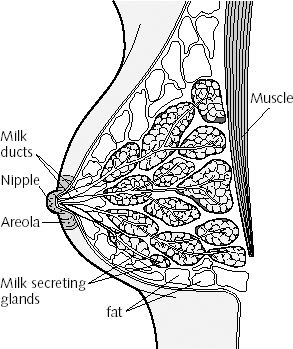
The breast is a mass of glandular, fatty, and fibrous tissues positioned over the pectoral muscles of the chest wall and attached to the chest wall by fibrous strands called Cooper’s ligaments. A layer of fatty tissue surrounds the breast glands and extends throughout the breast. The fatty tissue gives the breast a soft consistency.

The glandular tissues of the breast house the lobules (milk producing glands at the ends of the lobes) and the ducts (milk passages). Toward the nipple, each duct widens to form a sac (ampulla). During lactation, the bulbs on the ends of the lobules produce milk. Once milk is produced, it is transferred through the ducts to the nipple. 

The breast is composed of:
      *   milk glands (lobules) that produce milk 
      *   ducts that transport milk from the milk glands (lobules) to the nipple 
      *   nipple 
      *   areola (pink or brown pigmented region surrounding the nipple) 
      *   connective (fibrous) tissue that surrounds the lobules and ducts 
      *   fat


Brest Anatomy


The breast is a mound of glandular, fatty and fibrous tissue located over the pectoralis muscles of the chest wall and attached to these muscles by fibrous strands (Cooper's ligaments). The breast itself has no muscle tissue, which is why exercises will not build up the breasts. A layer of fat surrounds the breast glands and extends throughout the breast. This fatty tissue gives the breast a soft consistency and gentle, flowing contour. The actual breast is composed of fat, glands with the capacity for milk production when stimulated by special hormones, blood vessels, milk ducts to transfer the milk from the glands to the nipples and sensory nerves that give feeling to the breast. These nerves extend upward from the muscle layer through the breast and are highly sensitive, especially in the regions of the nipple and areola, which accounts for the sexual responsiveness of some women's breasts.  
 Because the breast is made up of tissues with different textures, it may not have a smooth surface and often feels lumpy. This irregularity is especially noticeable when a woman is thin and has little breast fat to soften the contours; it becomes less obvious after menopause, when the cyclic changes and endocrine stimulation of the breast have ceased and the glandular tissue softens. Estrogen supplements after menopause can cause continued lumpiness. The breast glands drain into a collecting system of ducts that go to the base of the nipple. The ducts then extend through the nipple and open on its outer surface. In addition to serving as a channel for milk, these ducts are often the source of breast problems


The ducts end in the nipple, (which projects from the surface of the breast), and are a conduit for the milk secreted by the glands and suckled by a baby during breast-feeding. There is considerable variation in women's nipples. In some, the nipple is constantly erect; in others, it only becomes erect when stimulated by cold, physical contact or sexual activity. Still other women have inverted nipples.

Surrounding the nipple is a slightly raised circle of pigmented skin called the areola. The nipple and areola contain specialized muscle fibers that make the nipple erect and give the areola its firm texture. The areola also contains Montgomery's glands, which may appear as small, raised lumps on the surface of the areola. These glands lubricate the areola and are not symptoms of an abnormal condition.

Beneath the breast is a large muscle, the pectoralis major, which assists in arm movement; the breast rests on this muscle. Originating on the chest wall, the pectoralis major extends from deep under the breast to attach to the upper arm. It also helps form the axillary fold, created where the arm and chest wall meet. The axilla (armpit) is the depression behind this fold.

Each woman's breasts are shaped differently. Individual breast appearance is influenced by the volume of a woman's breast tissue and fat, her age, a history of previous pregnancies and lactation, her heredity, the quality and elasticity of her breast skin and the influence of hormones

How to choose a Bra

How to choose a Bra


When choosing a bra for heavy breasts, make sure it has flat well-padded under cup wiring and wide elasticized straps that gives support to the centers of the breasts. Small breasts can be enhanced with the help of highly padded bras. Now the question is will the breasts sag, if you do not wear a bra? The sagging is due to slackening of the supporting muscles, and is more possible if the breasts are heavy. Wearing a well-fitting bra will delay sagging for little while. Always wear a good supporting bra during exercises, running, jogging and pregnancy. Exercising neither reduces the size of heavy breasts, nor do helps increase its size. Swimming is an excellent exercise for the bust shape and it strengthens the muscles. This is reason by swimmers have wonderfully shaped bust line.

During pregnancy and the period of breast-feeding, the breasts become larger and elastic fibres in the skin tear, showing red irregular marks on the skin surface. By regular massage with a moisturizing cream the elasticity of the skin is increased and the breast skin becomes soft and supple. Remember to remove bras at night. An underdeveloped breast is a matter of great concern for women, but massaging with a nourishing cream is the only remedy.
Choosing A Good Sports Bra


It is really important to wear a good sports bra when exercising regardless of the size of your bust. It will help to reduce breast pain and minimise 'sagging' in the future! But many women don't know how to choose the right style or size. Here are our tips for finding the right sports bra.

- Make sure that the bra is specially designed for sports use. Most bras are not designed for the increased movement during exercise. If you want to use a non-sports bra you already own, make sure it has a 'firm support'.

- Measure your size properly. Most of the women wear wrong size bras.

- Check the bra strap. It should be firm but comfortable. If it is too loose the bra won't stay put, but too tight and it will be hard to breathe.

- Check that the cups are the right size. If there is any bulging the cup size is too small, if the cups wrinkle the cup is too big. It is important to get the right cup size.
Supporting Your Breasts


Breasts have no muscles of their own and are only held up by the pectoral muscles—the muscles of the chest on which they lie. Extra support can be given by wearing a good bra. Most of the support that well-fitting bra gives to the breast should come from beneath and not from the straps. You can check this by slipping off the straps to see if the bra will stay in place without them. The back piece and the sides of a good bra should be in level with the front.How to measure the Bra Cups


If the bra cups are creased all over, then they are too big for your breast size. To find out the correct size of the bra cup, first measure around your rib cage under your breasts. Then measure around the fullest part of your bosom at the level of the nipple. The difference between the two measurements will give you the cup size.

How to use the Products and Steps for Massage


How to use the Products and Steps for Massage 


Left Breast Massage

1.) Position right hand below the armpit and at the same level of the breast and draw your hand inwards to the center of the body.

2.) Position left hand to the side of the breast following the contour of the breast with the thumb pointing outward and other fingers pointing downward, and push in from the side to the center of the body.

3.) Move the right hand slightly toward the center of the body, then use left hand to draw the breast from side and the base and massage in a clockwise motion.

* Repeat step 1 - 3 until the cream is fully absorbed.

Right Breast Massage

1.) Position left hand below the armpit and at the same level of the breast and draw your hand inwards to the center of the body.

2.) Position right hand to the side of the breast following the contour of the breast with the thumb pointing outward and other fingers pointing downward, and push in from the side to the center of the body.

3.) Move the left hand slightly toward the center of the body, then use right hand to draw the breast from side and the base and massage in a counter-clockwise motion.

Brest Health Tips


 Press Room

" There's new information that suggests an herbal plant will enhance your breast. News Channel 5 reports new finding show if women take an herb called Pueraria Mirifica; it will increase their breast size.

The root is presumed to contain substances that might have an effect on glands," say medicinal plant expert Vero Tyler, Ph.D. The herb is so popular that demand is outweighing supply." - NEWS NET 5

"Compared with women in the United States, women in Asia are far less likely to develop cancer of the breast or uterus. Many researchers believe that the differences in diet are one possible explanation. And on key component may be Pueraria Mirifica herb - plant compounds that have beneficial, hormone - like effects in the body." - Health News from the publishers of the New England Journal of Medicine "On Health" 

"In Thailand they have an herbal plant that is supposed to help in growing your breasts, and they are afraid it may fall into foreign hands. 

The Pueraria Mirifica root has been popular with Thai women for decades. The plant is supposed to have an estrogenic effect and can enlarge the breasts and hips of women." - Bangkok Post

"A jungle root that grows in the north of Thailand is believed to enlarge the breasts of women who take it. Firms from Japan, Germany and the United States have been conducting research into the Pueraria Mirifica root, a source in Thailand's Agriculture Department told the Bangkok Post. 

It has been used by Thai women for decades, Wichai Cherdshewasart of Chulalongkorn University in Bangkok told Reuters. 

"It shows an oestrogenic effect. It can enlarge breasts and hips. It will vary from woman to woman. If a woman's breasts are large already, the effect will be greater." - Bangkok Post


Brest World of the Body: breast


World of the Body: breast 


 The human breasts are mammary glands — common to all mammals, by definition. There are differences between species in number and in structure, and also in the composition of the milk that they produce for feeding the offspring.

A pair of nipples is of course common to both boys and girls — a relic of the embryological development of the male having been superimposed upon the basic female. As girls approach puberty, female sex hormones produced in the ovaries circulate in the bloodstream and cause development the rudimentary breast glands, which have been present since before birth. Breast enlargement usually heralds the other changes. Progesterone promotes development of the potentially milk-producing cells, and oestrogens promote the development of the ducts leading to the nipple from the 15-20 ‘lobes’ of glands.

When menstrual cycles begin, the mammary glands also start to undergo cyclical changes: in the second half of the cycle, under the increasing influence of progesterone, the glandular tissue grows, sometimes causing ‘lumpiness’ and tenderness — one of many preparations for the pregnancy which in most months does not follow.

When conception does occur, the breasts continue to develop. The accompanying increase in blood supply distends the veins under the skin — often the first outward and visible sign of pregnancy. The glandular tissue proliferates, taking the place of connective tissue and fat, and the breasts progressively enlarge. Later in pregnancy, the hormones secreted from the fetal tissue of the placenta act on the glandular cells and on the ducts leading to the nipple: thus the fetus itself, along with the hormone, prolactin, from the mother's pituitary gland, prepares the ground for its own later nutrition. This same prolactin would also stimulate the production of milk — but oestrogens from the placenta counteract this, so that milk is not actually made before the time is ripe. After birth of the baby, this suppression stops, so prolactin activity is suddenly uninhibited. Unfortunately for ideal infant feeding, in ‘developed’ countries nowadays oestrogens are often taken orally, to suppress milk production in those mothers who choose to bottle-feed the baby.


The female breast. After Youngson, Encyclopedia of family health


Left to nature, the secretion is initially scanty (colostrum) but the volume of milk becomes significant at about the third day after the birth, when the breasts become quite dramatically engorged. The mother's pituitary hormones remain in control of milk synthesis and secretion; under this influence, fats, proteins, and lactose (milk sugar) are made in the gland cells from nutrients taken up from the blood. When the infant sucks, nerve impulses from the nipple reach the hypothalamus in the brain; these stimulate nerve cells that have stores of the hormone oxytocin in the ends of their fibres that lie in the posterior part of the pituitary gland. This causes release of the hormone into the circulating blood. Reaching the breasts, oxytocin activates contractile cells, which squeeze milk from its storage sites into the channels that take it to the nipple. This whole ‘neuroendocrine reflex’ takes about 10 seconds — barely long enough for a hungry infant to show serious signs of frustration.

Lactation will continue for just as long as a baby is regularly sucking away the supply of milk: the more is removed, the more is made. The volume averages about 1 litre per day, but twice that amount can be produced for twins. Weaning of the infant leads automatically to a decrease in the milk supply, and the glandular tissue reverts to the non-pregnant state — until the next time, if any.


The breasts in history and culture

The cultural significance of the breast revolves around its uses as a symbol both of fertility and of sexual pleasure.

Many prehistoric images represent the female body with a high level of body fat and large breasts, the ideals when the food supply was uncertain. Breast milk, as our first and most reliable food, has long been the subject of speculation about its nature and significance. The classical model dominant in Western medicine until the nineteenth century was dependent both on the Greek philosopher Aristotle, who argued that breast milk was a fluid intermediate between menstrual blood and semen in terms of the degree of ‘cooking’ it received in the body, and also on the Hippocratic medical writers. It was thought that special channels from the womb to the breasts carried and transformed blood; this meant that, after birth, a child continued to derive nourishment from the same blood that had been its source in the womb.

The medical imperative from such theories was, of course, that a mother should nurse her own child. However, in cases where the natural mother was unable to do this, or as a way of preserving the youthful appearance of the breasts, wet-nursing could be used. Contracts specifying the duties of a wet nurse, and her fees, survive from Roman Egypt, showing that this form of paid employment was available to women from early times. The second-century ad medical writer, Soranos, offered detailed, and historically influential, advice to Roman men on how to choose a good wet nurse. She should be aged between 20 and 40, have given birth two or three times, and be strong and in good health. Her breasts should be medium-sized, soft, and unwrinkled, with the nipples also of medium size and neither too compact nor too porous. Soranos argued that milk from large women is more nourishing, but regarded very large breasts as a health risk to the infant on two counts: first, they may fall on the nursling, and second, there will be milk left over after each feed, which will lose its freshness and then harm the infant at the next feed. Soranos believed that the wet nurse transmits her own qualities to the child, so an even-tempered woman free from superstition should be found; she should also be Greek-speaking, so that the nursling becomes accustomed to hearing Greek. The wet nurse must abstain from sex and alcohol, both of which could damage the milk. As well as studying the body of the potential employee, a Roman man must taste and smell her milk; after employing her, he should carefully supervise her diet. In the nineteenth century the recognition of the value of colostrum superseded the classical view that, not being ‘proper milk’, it should be withheld from the baby.

The advice Soranos gives represents both a continuing unease surrounding the use of wet nurses, and a continuing conflict between the nurturing and the erotic breast. Roman writers often accused women of wanting to employ a wet nurse only for the sake of maintaining a sexually desirable figure. Medium breasts on large women may have been good for babies, but classical art suggests that the erotic ideal was the small breasted, boyish woman.

In mid-eighteenth-century Europe when Linnaeus' classification of the natural world put humans among the Mammalia — those with breasts — debate over the use of wet nurses became a state concern. Linnaeus was in favour of mothers nursing their own children; with philosophers, naturalists, moralists, and medical writers, he argued that strong nations were built up from babies fed at the maternal breast. Using the maternal breast was economical, but also political, part of the good woman's civic duty, and linked to images of the state feeding its children.

When, as a result of Pasteur's discoveries, sterilization of animal milk for bottle-feeding became possible, even those who could not afford to pay a wet nurse could avoid breastfeeding. A further development was milk substitutes; however, in developing countries there have been considerable problems following the promotion of milk substitutes as an alternative to the real thing, due for example to the formula being made up with non-sterile water or at the wrong strength.

The patron saint of nursing mothers is St Agatha, the legendary martyr who had her breasts cut off, shown in renaissance and baroque art carrying them on a plate. Christian religious art has used the nurturing breast in many ways. In fourteenth-century Tuscan art, during a period of crop failures and plague, the image of the Virgin Mary suckling a greedy Jesus became widespread. Sometimes she is shown directing a stream of milk into Jesus' mouth, or into the mouth of a particularly privileged saint; such images can emphasize the humanity of Jesus, or evoke the analogy of the Christian sucking at the breasts of the church for spiritual nourishment. Images of Charity personified often show a child suckling at each of her breasts.

For Freud, the breast was the first erogenous zone, from which a child should move on to the anal and genital stages of its developing sexuality. The baby's complete satisfaction at its mother's breast led to an identification with the mother, after which the baby needed to develop a sense of itself as a separate being. This was achieved by a rejection of the breast, now seen as withholding milk. In adult life, a person therefore longs for the perfect pleasure of the breast which has been taken away. Ideals and representations of the erotic breast show far more variation than the lactating breast. It can be large or small, with a pronounced cleavage or with the breasts entirely separated. The ideal in the Middle Ages was to have firm, white, apple-shaped globes, far from the Hollywood images of Jane Russell or Lana Turner, and even further from the pneumatic breasts of top-shelf magazines. Sixteenth-century kings' mistresses, most notably Agnès Sorel, Diane de Poitiers, and Gabrielle d'Estrées, were painted showing their breasts; Agnès was even represented as the Madonna.

As the size and shape of the ideal breast has varied dramatically over time and space, so fashions have changed to reshape the normal range of breasts to fit the ideal. The breast has been compressed, surgically reduced, padded, enhanced with silicone, pushed up, and armoured by a range of devices including bodices, corsets, bras and, most recently, the Wonderbra. Even before the corset or the brassiere, in the Middle Ages pouches sewn in to dresses could give uplift. One of the best-known aspects of the early Women's Liberation Movement was the ‘bra burning’ of the late 1960s, a form of liberation intended to make men face up to the reality of the breast freed from its fantasy underpinnings.

Breast tissue is more prone than any other in the woman's body to develop cancer. This accounts for about 1 in 20 deaths of British women, becoming commoner with increasing age. Early detection is assisted by regular X-ray examination (breast screening — mammography), and various combinations of surgery, radiotherapy, and chemotherapy can be effective in treatment.

The very high incidence of breast cancer in the Western world has made the breast into an organ associated as much with death as with nurturing life. Fanny Burney's harrowing description of her mastectomy, performed without anaesthetic in 1811, has survived; nowadays ‘lumpectomy’ may be adequate but mastectomy is sometimes necessary, and women who have had a breast removed may choose to use a prosthesis, or to adjust to a new body shape. The classical myth of the Amazons presents the woman with one breast as powerful, but feared. Currently some women with a family history of breast cancer are offered elective surgery to remove both breasts before disease appears; reactions to those who accept this surgery show that the breast remains a potent symbol of womanhood today.

— Sheila Jennett, Helen King

Sci-Tech Encyclopedia: Breast


 Sci-Tech Encyclopedia: Breast
Home > Library > Science > Sci-Tech Encyclopedia

The human mammary gland, usually well developed in the adult female but rudimentary in the male. Each adult female breast contains 15–20 separate, branching glands that radiate from the nipple. During lactation their secretions are discharged through separate openings at the base of the nipple.

In the female, hormonal changes in adolescence cause enlargement of breast tissue, but much of this is connective tissue although some glandular buds form. With the advent of full menstruation ovarian estrogenic hormones influence breast development. If pregnancy ensues, the glandular tissue reaches full development and full lactation begins shortly after birth. After cessation of lactation the breasts regress considerably and once again reflect cyclic regulation. See also Lactation.

Breast disorders may result from congenital or developmental abnormalities, inflammations, hormonal imbalances, and, most important, from tumor formation.

Congenital defects are usually unimportant except for their psychic or cosmetic implications. Supernumerary nipples and breasts or accessory breast tissue are common examples.

Inflammations are not encountered frequently and usually result from a staphylococcal or streptococcal invasion incurred during lactation. A special form of inflammation may result from fat necrosis. Although any age is susceptible, older women show a slightly higher incidence of fat necrosis, the commonest cause of which is injury from trauma. See also Staphylococcus; Streptococcus; Syphilis; Tuberculosis.

Hormonal imbalances are believed to be responsible for the variants of the commonest nontumorous breast disorder of women, cystic hyperplasia. The changes are thought to result from exaggeration or distortion of the normal cyclic alterations induced during the menstrual interval. Although a wide range of clinical and pathologic variation is commonplace, three major types or tendencies prevail. The first, called fibrosis or mastodynia, is marked by an increase of connective tissue in the breast, without a proportionate increase in glandular epithelium. The second, cystic disease, is characterized by an increase in the glandular and connective tissues in local areas, with a tendency toward formation of cysts varying in size. The third major type is adenosis, in which glandular hyperplasia is predominant. Each major form of cystic hyperplasia has its own clinical characteristics, ages of highest incidence, and distribution. Each is important because the breast masses which occur require differentiation from benign and malignant tumors. These lesions also have been found to predispose to the subsequent development of carcinoma.

Breast cancer is the most significant lesion of the female breast, accounting for 25,000–30,000 deaths in the United States each year. It rarely occurs before the age of 25, but its incidence increases each year thereafter, with a sharper climb noted about the time of menopause. Early breast cancer may appear as a small, firm mass which is nontender and freely movable. Diagnosis at this time carries a more favorable prognosis than later, when immobility, nipple retraction, lymph node involvement, and other signs of extension or spread are noted. Paget's disease of the nipple is a special form of breast cancer, in which there are early skin changes about the nipple

S I T E

http://www.GetMonthlyPay.com/index.php?invite=54667

Visitors

free counters

About This Blog

Lorem Ipsum

Usefull Link

  © Blogger templates Shiny by Ourblogtemplates.com 2008

Back to TOP